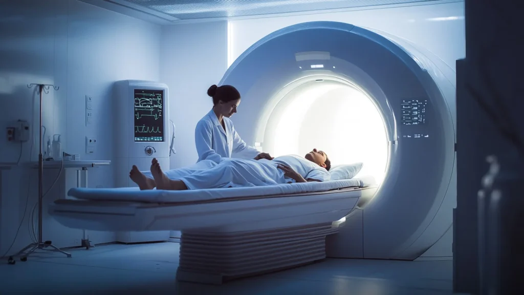
In our modern-day, there exist several reliable imaging diagnostic systems: X-rays, ultrasound, MRI, CT, and PET scans. When a patient’s symptoms indicate a possible growth or significant organ dysfunction, medical professionals will most likely want to use one or more of these systems to verify how serious the problem is.
As a patient, you may be concerned as to how safe these systems are. In the case of X-rays, CT, and PET scans, you are probably aware that these scans expose your body to radiation. What are the risks of radiation, how much radiation is your body exposed to during these scans, and what are some things you need to bear in mind?
What is ionizing radiation?
The World Health Organization (WHO) says: “Ionizing radiation is radiation with enough energy so that during an interaction with an atom, it can remove tightly bound electrons from the orbit of an atom, causing the atom to become charged or ionized.”1
There are two types of electromagnetic waves that can ionize atoms: X-rays and gamma-rays.
Put simply, Ionizing radiation is any type of particle or electromagnetic wave that carries enough energy to ionize or remove electrons from an atom. Once these atoms begin to decay they expel energy, which is so strong it negatively affects other molecules that lay in its path.
How much radiation are we being exposed to during medical scans?
Ionizing radiation is measured in millisieverts (mSv). Taking a 10-hour flight, for example, can expose you to around .03 mSv of ionizing radiation and a dental X-ray is approximately 09 mSv. However, this level of exposure increases dramatically for other forms of scans.
- A mammogram X-ray exposes a woman to .4 mSv.
- A chest CT gives off 8 mSv.
- An abdominal CT scan uses 10 mSv.
- Most PET scans expose you to 14 mSv.
As a precaution, those working in radiation-related occupations, such as radiographers, must not be exposed to more than 50 mSv per annum.
Anyone potentially at risk or already suffering from cancer may be concerned whether undergoing such diagnostic procedures will put them at more risk.
Medical professionals are reliant on medical imaging scans. The general approach is that the benefits of such scans far outweigh the risks. Unfortunately, the imaging process is not the only exposure to radioactivity that some patients are subject to. In order to provide a full picture, some patients will need to inhale, swallow, or have radioactive materials injected into them as part of the diagnostic process.
Positron Emission Tomography (PET) scans, for example, work by using a radioactive tracer, which may be taken orally or injected depending on the particular organ being studied. In the case of injections, glucose tagged with radioactive material enters into the body intravenously. The glucose goes straight to the cancer cells and the radioactive material lights up the scan, showing the radiographer where the cancer is, how big it is, and its SUV (Standardized Uptake Value).
Naturally, doctors advocate these scans because they deliver a clear picture of what is happening within a patient’s body. To them, these scans are well worth any potential risks, if they allow them to obtain a correct diagnosis in order to determine the necessary treatment at the earliest possible stage. As a patient, you may also agree with this way of thinking. Remember that you always have a choice of whether to accept or decline any scan that is offered or recommended. The ultimate purpose of a scan is to obtain an image of some part of your body. Questions you may ask yourself or your doctor before accepting a scan are:
- For what reason is this scan necessary?
- What are the risks involved?
- Is there a less harmful scan that could be conducted instead? Is there a way to conduct the scan in a way that would expose my body to a lower amount of radiation?
- If the scan confirms that there is a tumor, what course of action or treatment will be recommended by my doctor?
- If the scan shows that the tumor has grown since the last scan, what will be recommended and what do I plan on doing?
Preparing yourself ahead of time and having a clear idea of your and your doctor’s purpose for conducting any given scan may help you to make a balanced decision
What are your options?
You can probably see why a PET scan is an invaluable investigatory method for doctors. At the time of writing this, there are, unfortunately, no other tests that can measure metabolic and chemical functions in the body in the same way a PET scan can do. So, it is a case of weighing up the pros and cons for yourself as a patient. It is imperative to remember that you can, and should, say no to tests that you feel are unnecessary.
If possible, ask for non-radiative tests such as MRI and ultrasounds first.
If you must have a PET scan, you can try to minimize the risks by asking for the following:
- A shield to protect parts of your body that aren’t being imaged
- The lowest possible dose of radiation
- That your technician pays attention and uses as little radiation as possible for the shortest possible time.
- To be referred to a newer machine that uses lower radiation.
After the scan
Once you have had your scan, you can use food therapy to proactively protect your DNA. Eating foods like mushrooms, avocado, applesauce, garlic, lemons, seaweed, elderberry syrup, and miso are great for their essential fatty acids.
Certain plants and herbs have shown to have radioprotective properties. Some are listed here:
Gingko Biloba, Centella Asiatica, common sea-buckthorn (Hippophae rhamnoides), holy basil (Ocimum sanctum), ginseng (Panax ginseng), Himalayan mayapple (Podophyllum hexandrum), amaranth (Amaranthus paniculatus), Indian gooseberry (Emblica officinalis), Phyllanthus amarus, long pepper (Piper longum), heart-leaved moonseed (Tinospora cordifolia), mint (Mentha arvensis), peppermint (Mentha piperita), jambolan (Syzygium cumini), ginger (Zingiber officinale), billygoat-weed (Ageratum conyzoides), bael (Aegle marmelos) and Aphanamixis polystachya.2
You could also try dimethyl sulfoxide (DMSO). A Japanese study showed even low doses of DMSO had full-body radioprotective effects.3
It is important to remember that YOU are your own best advocate. Empowering yourself with knowledge in advance of any doctors’ visit can help you to understand all the risks involved and weigh up the pros and cons. ALWAYS ask what alternative options are available to you and take the time to research before committing to anything. Of course, a speedy diagnosis in the case of cancer gives patients the best possible chance of recovery, but it is important to ensure that you are not putting yourself at any unnecessary risk.
It is important to have a clearer understanding of what is happening in the body, the cause of the imbalance, as well as a solution, and that is what we are here for. Our Cancer Home Care Program gets to the root causes of cancer and addresses them.
References:
- What is Ionizing Radiation? – WHO – World Health Organization:http://www.who.int/ionizing_radiation/about/what_is_ir/en/
- Radioprotective Potential of Plants and Herbs against the Effects of Ionizing Radiation – National Center for Biotechnology Information, U.S. National Library of Medicine: https://www.ncbi.nlm.nih.gov/pmc/articles/PMC2127223/
- An alternative mechanism for radioprotection by dimethyl sulfoxide; possible facilitation of DNA double-strand break repair – National Center for Biotechnology Information, U.S. National Library of Medicine: https://www.ncbi.nlm.nih.gov/pubmed/21116101